syngo DynaCT

syngo DynaCT, introduced in 2004, is Siemens’ cone-beam CT solution enabling users to obtain soft-tissue information during interventions. It gained recognition as a highly valuable tool for numerous clinical procedures. syngo DynaCT improves patient outcomes by providing information not apparent in DSA, and allows users to broaden their procedure scope. Being perceived in the past as more suitable for expert users, the new PURE® platform now makes it easier for all users to achieve consistent high-quality results.

  • Support consistent results in cone-beam CT
  • Improve patient outcomes and avoid complications
  • Broaden the procedure scope of your angio lab

Características e Benefícios

syngo DynaCT enables CT-like cross-sectional imaging by creating 3D soft tissue data sets. syngo DynaCT can be performed directly in the angio suite without loss of time and additional risk to the patient. 

In the past, different levels of expertise may sometimes have led to varying results when using cone-beam CT. In contrast, with the new 3D Wizard image-guided protocol selection, available on Artis with PURE®, syngo DynaCT is also accessible for occasional users. The 3D Wizard leads all users to consistent and high-quality results. It helps users select the correct protocol with a visual support of clinical images, offers all relevant information on delay, injection and patient positioning, and guides users through the acquisition, step by step. This supports standardized quality of care, regardless of who performs the procedure, from occasional to experienced users.

Improve patient outcome

syngo DynaCT can provide information not apparent in DSA. This additional information may result in changes in diagnosis, treatment planning, or treatment delivery. It supports sounder decision-making during the intervention – for example when employed to exclude non-target embolization during liver tumor embolization or to confirm device position within intracranial vessels. Where in the past it would have been necessary to transfer the patient into the CT unit, syngo DynaCT can be performed instead, directly in the angio suite without loss of time and with no additional risk to the patient.

Broaden the procedure scope of ypu angio lab

Dedicated syngo DynaCT applications allow users to extend the clinical capabilities of their angio lab. syngo DynaCT Micro, for instance, enables users to visualize the smallest details, bringing additional procedures such as arthrography, intra-cranial stent placement follow-up examinations, or validation of cochlear implant placement to the angio suite. Other dedicated applications that further expand the confidence and performance of syngo DynaCT include syngo DynaCT SMART (Streak Metal Artifact Reduction Technique): This special technique, available for Artis with PURE®, substantially reduces metal artifacts, enabling sounder detection of bleeds even around small coil packages. syngo DynaCT 360 or syngo DynaCT Large Volume: Enable acquisition of images with an even larger volume, which can be very useful during liver embolization or needle procedures

Employ your workforce optimally

syngo DynaCT also enables users to take on recently developed, innovative procedures – thus extending their clinical capabilities even further. For instance, in new embolization procedures such as prostate artery embolization for treatment of benign prostatic hyperplasia, syngo DynaCT helps operators to clearly distinguish between target and non-target embolization sites. syngo DynaCT gives users the right tools to tackle exciting new prospects.

Uso Clínico

Siemens was first to introduce soft tissue imaging in the interventional suite as a breakthrough innovation for neuro applications. Continuous development in collaboration with institutions worldwide has made syngo DynaCT available for applications in the entire body. What started as a breakthrough is now a worldwide trend in interventional imaging. As of June 2015, hundreds of institutions around the world have installed syngo DynaCT in their angio suite.


Minimally Invasive Spinal Osteosynthesis

Minimally Invasive Spinal Osteosynthesis
An 88-year-old female patient suffered from lower back pain. She was operated on before due to a traumatic fracture of L5 which required cementoplasty.

Stent Placement across Cerebral Aneurysm

Stent Placement across Cerebral Aneurysm
65-year-old male with hypertension and history of myocardial infarction and coronary stenting with incidental left internal carotid artery aneurysm.

Cerebral trauma

Cerebral trauma
Female patient with brain trauma for 3 days, mental imbalance and loss of consciousness.


syngo DynaCT in multiple phases for HCC

syngo DynaCT in multiple phases for HCC
A 69-year-old male with HCC (hepatocellular carcinoma) and HCV (Hepatitis C virus)-positive hepatic cirrhosis.

Follow-up after stent-assisted aneurysm coiling

Follow-up after stent-assisted aneurysm coiling
73-year-old female patient. Left middle cerebral artery (MCA) aneurysm that was treated with stent-assisted coil embolization in 2005. Because of her history of contrast allergies, we decided to complete a syngo DynaCT run with IV injection...

syngo DynaCT of the pulmonary arteries

syngo DynaCT of the pulmonary arteries
Patient with history of recurrent pulmonary embolism and chronic thromboembolic pulmonary hypertension (CTEPH). Pulmonary angiogram and syngo DynaCT were acquired for diagnostic work-up.


syngoDynaCT Whitepaper 2015

Use of Cone Beam CT (CBCT) in the angio suite improves safety and outcomes in interventional tumor treatment

Dr. Michael J. Wallace

Dr. Michael J. Wallace on the Impact of Improved Image Quality of Angiography Equipment in Interventional Oncology


Detalhes técnicos

How does syngo DynaCT work?

syngo DynaCT utilizes images acquired from a special rotational angiography run to reconstruct cross-sectional soft tissue information. Image acquisition is achieved in 5-20 seconds, depending on the protocol used. In that time the C-arm covers an angle of at least 200° and acquires the needed projection images for reconstruction. These images are automatically sent and reconstructed on a syngo X Workplace and available for assessment in the interventional suite.

What does “soft tissue information” mean?

Before the introduction of syngo DynaCT, 3D angiography enabled the visualization of high-contrast objects such as stents, clips, bones and contrast-filled vessels during interventional procedures. With syngo DynaCT, it is possible to achieve a higher level of tissue differentiation in the reconstructed images, enabling the visualization of objects with lower Hounsfield Units (HU) values.

syngo DynaCT Improve

With syngo DynaCT we can visualize objects of 5 HU and 10 mm in size or 10 HU and 5 mm in size*. This enables visualization of lower contrast objects such as larger hemorrhages and tumors.
* as measured using a 16-cm CATPHAN phantom

What is the FOV (field of view)?
syngo DynaCT datasets acquired with a ceiling, floor or biplane stands provide a volume of 18.5 cm height with a diameter of 24 cm.
The Artis zeego enables in addition larger volume acquisitions such as syngo DynaCT Large Volume and syngo DynaCT 360, which result in a volume of 18 cm height with a 47 cm diameter, or 24 cm height with a 35 cm diameter.

syngo DynaCT FOV

If desired, it is also possible to acquire a smaller volume using collimation* in order to reduce dose and increase image quality.
*collimation on the longitudinal access.

What is the spatial resolution of syngo DynaCT and what slice thickness can be reconstructed?
The slice thickness that can be reconstructed depends on the volume of interest and the voxel size. The standard slice thickness is about 0.5 mm and there are additional reconstruction options that allow creation of lower slice thickness. The spatial resolution achieved with syngo DynaCT is about 0.22 mm. In addition, with syngo DynaCT Micro, a special protocol is available that provides high resolution 3D imaging, boosting the level of detail by using each detector pixel. With syngo DynaCT Micro an even higher spatial resolution of about 0.14 mm can be achieved.


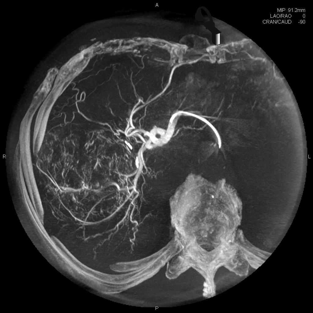
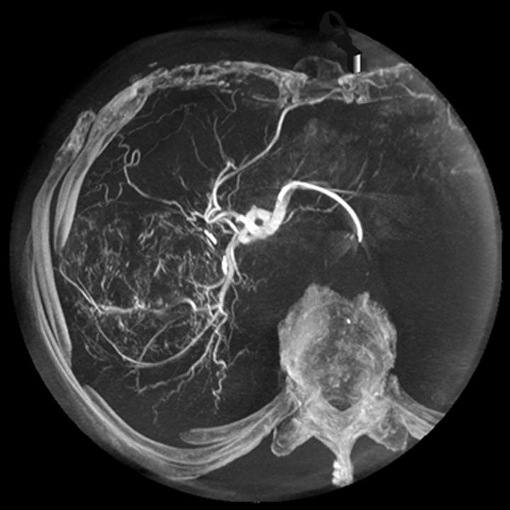
Does syngo DynaCT offer a dedicated body protocol?

Yes, syngo DynaCT offers a dedicated body protocol, about 400 projections are acquired in less than 6 seconds*. Due to its fast rotation this protocol reduces motion artefacts providing excellent image quality. High resolution results (512 matrix) are available at tableside in less than 15 seconds*. In addition there is a dedicated syngo DynaCT protocol with reduced dose and shorter acquisition time for dose-sensitive applications.
*Mentioned values are for Artis with PURE and may change depending on system hardware and specific protocol used.

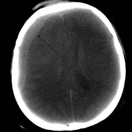
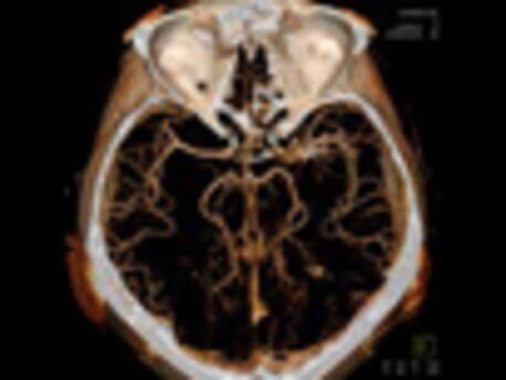
Does syngo DynaCT offer a dedicated neuro protocol?
Yes, there are several syngo DynaCT protocols that can be useful for interventional neuroradiology procedures.
The standard dedicated neuro protocol acquires about 500 projections in 20 seconds. High resolution results (512 matrix) are available at tableside in about 30 seconds*.
There are also protocols for reconstructing information of subtracted scans enabling the easy visualization of vessels in relation to devices such as stents or coil packages and a special protocol for stent follow-up using an intravenous injection.
In addition, with syngo DynaCT SMART (Streak Metal Artifact Reduction Technique) a special reconstruction is included for reducing metal artifacts in the resulting images, which increases the diagnostic confidence and the chance to visualize complications such as bleeds close to metallic objects.
* Mentioned values are for Artis with PURE and may change depending on system hardware and specific protocol used.

General Requirements

  • ARTIS pheno
  • Artis Q
  • Artis Q.zen
  • Artis zee

syngo X Workplace: VB21

Please note: Additional technical prerequisites may apply. Upon receiving your request, your local Siemens representative will clarify whether your system meets the requirements.

1